What Shape Are Cheek Cells
Cell cheek animal structures Cheek cells 400x stained Cheek microscope 40x onion nicholas
Amazing 27 Things Under The Microscope With Diagrams
Cheek human cell cells microscope methylene blue dye under bacteria skin microscopic stains dna mrc www2 toxic when experiment compound Solved using this table from the size estimation module, Cheek cell cells human animal membrane plant eukaryotic epithelium squamous lab cytoplasm post ppt powerpoint presentation obvious nuclei
Cheek cells 100x stained
Difference between onion cell and human cheek cell – pediaa.comCheek cells type microscopy enotes mag image007 further reading Cell structures & functionCells light onion viewed microscope cell human cheek bacteria bacterial biology shape skin electron nucleus size introduction micrograph rectangular nasal.
Cheek cellsDiagram of human cheek cell and onion cell Cheek cell human temporary stained prepare cells mounts microscope under lab observation work table shapeCells cheek human microscope 40x scp cell under 1809 stained 400x magnification blue swab total microscopic stain unstained thf biological.

Animal cheek cell under microscope
Cheek cell human diagramLesson 2: mount a slide & “look at your cheek cells“ Cheek cells microscope rsscience osmosis dictionary differently react biologyCheek methylene membrane microscope look stains bacteria biological organelles rsscience rod.
Cheek cells 100x human stainedMy opera is now closed Cheek cell lab – hailey's blogCheek cell microscope.
What type of cells are cheek cells?
Cheek cell bacteria cells human nucleus membrane using bacterial single been prokaryotic solved determineCheek cell under 40x 400x magnification cells lab nucleus nose piece Human cheek cells under the microscopeTo prepare stained temporary mounts of human cheek cell.
Cheek cells epithelialCheek cells are made up of(a)muscle cells(b)epithelial cells(c)nerve Cell cheek shape elodea expressions optics molecular scienceCell onion cheek between difference human comparison pediaa.
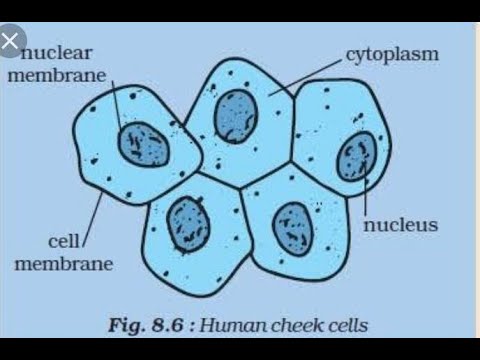
Cell onion cheek human diagram diagrams
Cheek cell cells onion 400x stained lab human animal slide biology staticflickr c1Diagram of. cheek cell Cheek cells lab – nicholas's blogCheek cells.
Cheek biologycorner cellsHuman cheek Cheek microscopic buccal meyer microscopesCheek microscope cell cells under human biology dna science banana shows pic swab hubpages part lesson each pearltrees 400x big.

Amazing 27 things under the microscope with diagrams
Cheek microscope theory 400x beings biologycorner timetoastEasy diagram for human cheek cell.....by tejbir mand... Microscope under cheek cells cell buccal nucleus lab other visible look staining theseWhat is inside your cheek?|| prepare the slide of human cheek cell.
Cells cheek microscope human under cell do animal membrane epithelium😊 shape of cheek cell. difference between onion cell and human cheek .


