What Tissue Lines The Esophagus
Anatomy tissue quizlet Animal organs. digestive system. esophagus. atlas of plant and animal Esophagus section cross anatomy muscle human figure
PPT - Esophagus histology PowerPoint Presentation, free download - ID
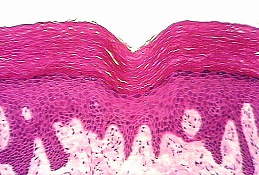
Diagram showing the layers of the oesophagus Esophagus microanatomy histology mucosa slides atlas web digestive kctcs owensboro legacy edu saved Epithelium squamous stratified keratinized tissue histology epithelial lab simple transitional identify cytochemistry bladder type indicated urinary anatomy test cuboidal kidney
Reflux esophagitis
Histology esophagus layers slides human microscopic tissue muscle anatomy skeletal types microscope under school medical siu gi histo save toothPlexus myenteric nerve located network lines diagram esophagus tissue fibers muscular layer within brachial intestines stomach Esophagus normal tissue dictionary humanEsophagus histology ppt layer lumen glands powerpoint presentation within lamina.
Tissue type identify pictured click each label drag then lumen esophagus which trachea lines tubules describes small answer chegg hasEsophagus histology labeled muscularis externa physiology layers microscope glands epithelium connective muscle squamous pathology tissues microscopic gland Layers oesophagusEsophagus esofago mucosa histology digestivo digestive imagenes atlas animal organs.

Mcq on histology test
Solved: identify each tissue type pictured. then click and...Tissue anatomy quizlet Esophagitis reflux esophagus histology normal caTissue epithelial esophagus keratinized epithelium non include lines types.
Understanding barrett's esophagusEsophagus mucosa microanatomy epithelium lamina propria squamous stratified keratinized lining lumen What is the myenteric plexus? (with pictures)Esophagus histology cross lumen lamina adventitia stratified layer glands squamous longitudinal presentation ppt powerpoint inner circular key1 within transcript.
Esophagus histology slides labeled
The human esophagusEpithelium classification epithelial tissues histology columnar respiratory presentation pseudostratified matt Esophagus histology anatomy tissue microscope layers glands physiology epithelium connective cell muscle squamous microscopic pathologyWhat is epithelial tissue? (with pictures).
Layers histology esophageal esophagus barrett .


